Reviews and articles
05.12.2018

Interpretation of RDW-SD and RDW-CV values in the diagnosis of anisocytosis
Very often, clients of laboratories where blood tests are performed on automatic hematology analyzers ask for clarification of the results obtained for all parameters of the general blood test. The question is natural, because instead of the usual five indicators, patients receive a printout, which contains from 18 to 22 indicators. Product Manager of the company "Diameb Trade" Svyatoslav Polovkovych brings to your attention an article that will help doctors and laboratory specialists to clarify this issue to patients.
Blood, its functions and what does “erythrocytes” mean
Blood is one of the important components of a living organism. It has the form of liquid tissue consisting of plasma and formed elements. By formed elements, we mean platelets, erythrocytes and leucocytes.
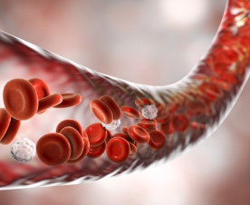
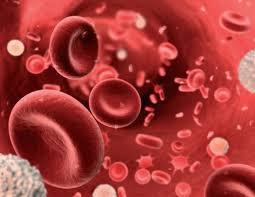
Erythrocytes or "red blood cells" (RBC) are nuclear-free cells that have a concave disc shape. The absence of a nucleus in erythrocytes and their shape provide them with the most optimal properties in the process of gas exchange and maintaining osmotic resistance. The normal size of the erythrocyte is 7.5-8.3 µm; life expectancy is 90-120 days. Red blood cells have antigenic properties, based on which four main blood groups are distinguished. The cytoplasm of erythrocyte by 96% filled with hemoglobin. In addition to mature erythrocytes, normally, young erythrocytes - reticulocytes - can be found in the peripheral blood. These are nuclear-free cells with a large number of RNA and ribosomes, which have membrane receptors for transferrin. In the reticulocyte stage, up to 30% of the total hemoglobin in the erythrocyte can be produced. Another 70-80% of hemoglobin is synthesized earlier, in the pre-reticular cell differentiation stages. Reticulocyte loses RNA and the ability to produce hemoglobin when it turns into a mature erythrocyte. In the reticulocyte stage, the erythrocyte exists in the bone marrow for one day and another day - in the peripheral blood.
The circulatory system connects and nourishes all organs, so it is very important to monitor its condition and regularly perform a general blood test. When conducting a general blood test should pay attention to the size, color and shape of blood cells. Poikilocytosis and anisocytosis can be considered based on the deviation of the shape and size of cells from normal values.
Signaling message of hematological analyzer "Anisocytosis":
what indicators to pay attention to
Specialists of laboratories often have to answer the question on the interpretation of hematology analyzer indicators. Automatic hematology analyzer itself informs about the clinical picture of the patient. However, the most asked questions at meetings of laboratory assistants are questions on the basis of what indicators these conclusions are made, and questions of interpretation of hematology analyzer indicators. Let us try to explain these aspects using the example of the interpretation of the values of the RDW-SD and RDW-CV indicators in the diagnosis of anisocytosis.
Blood anisocytosis is the excess of the number of cells of non-standard size. Depending on which formed elements of blood have changed their size, anisocytosis of erythrocytes and platelet anisocytosis are distinguished.
Erythrocyte anisocytosis in general blood test suggests that the size of blood particles differs from the standard. The normal size of erythrocytes is 7.5-8.3 µm. The presence in the blood of a small number of red blood cells of a non-standard size (compared to the total) is allowed. On average, this value is 30%. It is considered normal if in the blood 15% of cells are smaller than standard cells, and 15% of cells are larger. Erythrocytes with a smaller diameter (< 6.9 µm) are called microcytes. Erythrocytes with a large diameter are divided into two groups:
- macrocytes: 8 µm < d < 12 µm;
- megalocytes: d > 12 µm.
If the blood cells differ significantly in size from the allowable value, the patient can be diagnosed with «Increased erythrocyte anisocytosis".
Based on the size of the cells that prevail in the blood, microcytosis, mixed type and macrocytosis are distinguished. Mixed-type anisocytosis is intermediate between microcytosis and macrocytosis; this type is characterized by the presence in the blood of both small and large blood cells. For example, mixed-type anisocytosis with a predominance of microcytes means that the particle sizes in the blood are not uniform, but the majority are small particles.
To indicate the degree of heterogeneity, there is a special index - RDW (Red cell Distribution Width) or erythrocyte anisocytosis index. That is, the RDW demonstrates the inhomogeneity of the erythrocyte population size in the study sample.
There are two types of indicators - RDW-CV and RDW-SD. The first, RDW-CV, shows the percentage distribution of cells by size. The second, RDW-SD, is their standard deviation, that is, the difference in size between the smallest and largest erythrocyte in a blood sample.
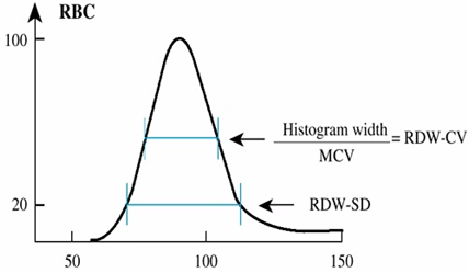
A hematology analyzer determines using a special formula that takes into account mean corpuscular volume of erythrocytes (MCV) and the standard deviation from MCV, the RDW-CV index automatically. The indicator calculated in this way is expressed as a percentage. The rate of RDW-CV is 11-15%. RDW-CV directly depends on the value of the corpuscular volume of erythrocytes (MCV), therefore, if the majority of cells in the population are small (as in microcytosis), the RDW-CV will remain within the normal range. RDW-CV is less sensitive to the presence of a small population of microcytes, macrocytes or reticulocytes, but better reflects the overall changes in the size of erythrocytes during macrocytic or microcytic anemia.
Also in the results of the blood test can be found indicator RDW-SD. This indicator is determined by a different method and does not depend on the corpuscular volume of erythrocytes. The determination of RDW-SD is a direct measurement of the width of erythrocytic histogram at 20% of the height of the curve; the height of the RBC histogram is taken as 100%. RDW-SD is measured in fl (femtoliters) and reflects the difference between the maximum and minimum volume of erythrocytes in the study sample. Normally, the RDW-SD is 35-60 fl. RDW-SD is a more sensitive indicator when a small number of macrocytes and microcytes appear in the erythrocyte population, since it measures the lower part of the erythrocyte distribution by volume. In addition, this indicator will change more quickly with reticulocytosis, since there will be a broadening of the base of the erythrocytic histogram.
The clinical value of RDW in the diagnosis
Interpretation of the RDW value in test results is always carried out in parallel with the valuation of the mean corpuscular volume of erythrocytes (MCV), since quite often the red cell distribution width remains normal in the presence of a homogeneous cell population during microcytosis or macrocytosis.
If the RDW is elevated in the blood test, the patient can assume the following pathological conditions:
- Iron deficiency anemia
- Hemolytic anemia of an immune nature
- Megaloblastic anemia (with a deficiency of vitamins B9 and B12)
- Hemoglobinopathy
Elevated values of RDW are also characteristic of patients with liver diseases and patients undergoing blood transfusions. In addition, the anisocytosis index may be overestimated erroneously if cold agglutinins are present in the study sample. It is also worth noting the pathologies in which RDW does not change. These include beta-thalassemia, anemia in severe chronic diseases, sickle cell anemia, acute hemorrhagic and aplastic anemia, and spherocytosis.
It is important that the study on an automated hematology analyzer is more accurate, since RDW can grow even before the appearance of changes in the red blood cells, and the hemoglobin value, that is, an increase in the anisocytosis index, can be called an early marker of anemia. It should be noted that during the treatment of iron deficiency anemia, the RDW indicator not only does not decrease, but also increases, and the histogram changes noticeably (two peaks appear on the distribution curve). This is due to the fact that there are young cells that differ in size from mature erythrocytes. If drug therapy is effective, anisocytosis index is normalized, but it normalizes the latest of all red blood cell indices.
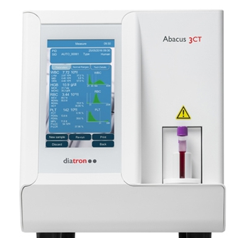
The "Diameb Trade" company offers automatic hematological analyzers of the European manufacturer the DIATRON Company. These analyzers measure, in addition to other standard indicators, the red cell distribution width (RDW) in two variants: RDW-CV and RDW-SD, which fully shows the inhomogeneity of erythrocyte population sizes in the sample under study. A wide range of DIATRON analyzers is represented by analyzers of various capacities (from 30 to 80 tests per hour), with different technical characteristics (small sample module, auto puncture function, autoloading, etc.). This allows you to fully meet the needs of laboratories of various sizes and directions (primary care health centers, family doctors, pediatrics, oncology, etc.). The company offers an individual approach to each client under the terms of payment, delivery and service. We provide warranty and after-sales service.